You closed it well.
Clean edges. Proper tension. Good apposition.
Then 3 to 5 days later…
Swelling. Fluctuance.
And suddenly, your incision is not holding.
That’s seroma.
WHAT IS A SEROMA?
A seroma is a sterile accumulation of serous fluid within a dead space created by tissue disruption.
It is not pus.
It is not infection.
It is plasma ultrafiltrate, inflammatory exudate, and lymphatic leakage that collects where tissues fail to adhere.
In small animal surgery, it is most commonly seen after:
- Extensive soft tissue dissection
- Mastectomy chains
- Flank spays
- Orthopedic procedures with large dead space
- Any surgery with poor tissue apposition
WHY DOES IT FORM?
Seroma formation is mechanical and physiologic.
Core mechanisms:
- Dead space → fluid has somewhere to accumulate
- Shear forces → disrupt fibrin bonds between tissue planes
- Lymphatic damage → continuous leakage
- Inflammatory response → capillary permeability increases
- Motion → prevents adhesion and promotes fluid buildup
This is why active dogs, obese patients, and poorly immobilized surgical sites are at higher risk.
WHY SEROMA LEADS TO DEHISCENCE
A seroma is not just fluid. It is pressure + separation + delayed healing.
1. Mechanical Tension from Within
Fluid accumulates and pushes tissue planes apart.
- Sutures are designed to hold approximated tissue, not resist internal expansion
- Increasing pressure converts a low-tension closure into a high-tension environment
- Eventually, something gives
2. Loss of Tissue Apposition
Healing requires direct contact between tissue layers.
- Fibroblasts cannot bridge the gap
- Fibrin scaffold formation is disrupted
- Angiogenesis is impaired
No contact = no healing = weak incision.
3. Compromised Perfusion
As fluid accumulates:
- Local pressure compresses microvasculature
- Oxygen delivery drops
- Tissue becomes hypoxic and fragile
Ischemic tissue fails faster under stress.
4. Inflammatory Amplification
Persistent fluid:
- Sustains inflammation
- Delays transition to proliferative phase
- Weakens collagen deposition
You end up with a prolonged inflammatory wound, not a healing one.
5. Increased Risk of Secondary Infection
While seromas are initially sterile:
- Fluid becomes a perfect medium for bacteria
- Even minor contamination can convert it to an abscess
Infection further degrades tissue strength and accelerates dehiscence.
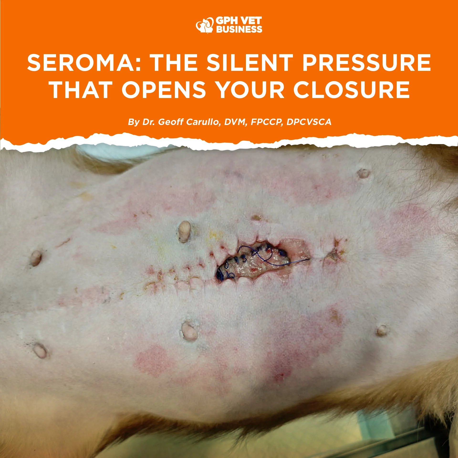
CLINICAL PATTERN YOU’LL RECOGNIZE
- Soft, fluctuant swelling near incision
- Non-painful initially
- No heat or redness (early stage)
- Appears 3 to 5 days post-op
- Incision edges start to stretch or separate
Then:
- Partial dehiscence
- Or full wound breakdown
PREVENTION IS TECHNIQUE
Seroma is often a surgical decision problem.
Reduce dead space:
- Walking sutures
- Layered closure
Minimize trauma:
- Gentle tissue handling
- Sharp dissection over blunt tearing
Control motion:
- Proper bandaging when indicated
- Strict activity restriction
Consider drains (when justified):
- Especially in large dissections or mastectomies
MANAGEMENT PRINCIPLES
- Small seromas → often self-limiting
- Large seromas → may require aspiration (with caution)
- Recurrent cases → address dead space, not just fluid
- Signs of infection → escalate immediately
Never treat the fluid alone. Treat the reason it exists.
REALITY IN PRACTICE
Most dehiscence cases blamed on:
- “makulit na aso”
- “hindi nag cage rest”
Many started as uncontrolled seromas.
Not all wound failures are behavioral. Some are architectural.
CLOSING LINE
A clean incision does not guarantee a stable one.
If you leave space…
the body will fill it.
And sometimes, that fluid is what breaks everything apart.
SOURCES
- Pavletic MM. Atlas of Small Animal Wound Management and Reconstructive Surgery, 4th ed. Wiley-Blackwell.
- Fossum TW. Small Animal Surgery, 5th ed. Elsevier.
- Swaim SF, Henderson RA. Small Animal Wound Management, 3rd ed.
- Johnston SA, Tobias KM. Veterinary Surgery: Small Animal, 2nd ed. Elsevier.
- Williams J, Moores A. BSAVA Manual of Canine and Feline Wound Management and Reconstruction, 2nd ed.
Dr. Geoff Carullo is a Fellow and the current President of the Philippine College of Canine Practitioners.
Sharing this helps others understand what it really means to be a vet. If you found this valuable, like and follow for more insights.