What if veterinarians could actually watch what is happening inside a patient’s body instead of relying on a single X-ray image?
Imagine watching a dog’s trachea collapse during breathing, observing how food travels through the esophagus, or guiding a catheter through a tiny blood vessel without making a large surgical incision.
This is no longer science fiction.
Welcome to veterinary fluoroscopy, where X-rays become moving images.


WHAT EXACTLY IS FLUOROSCOPY?
Traditional radiography produces static images. It allows veterinarians to examine bones, organs, foreign bodies, and abnormalities at a particular moment.
Fluoroscopy takes diagnostic imaging a step further.
Using a continuous or pulsed X-ray beam, it produces real-time moving images of internal anatomical structures.
Think of conventional radiography as taking a photograph.
Fluoroscopy is like watching a video.
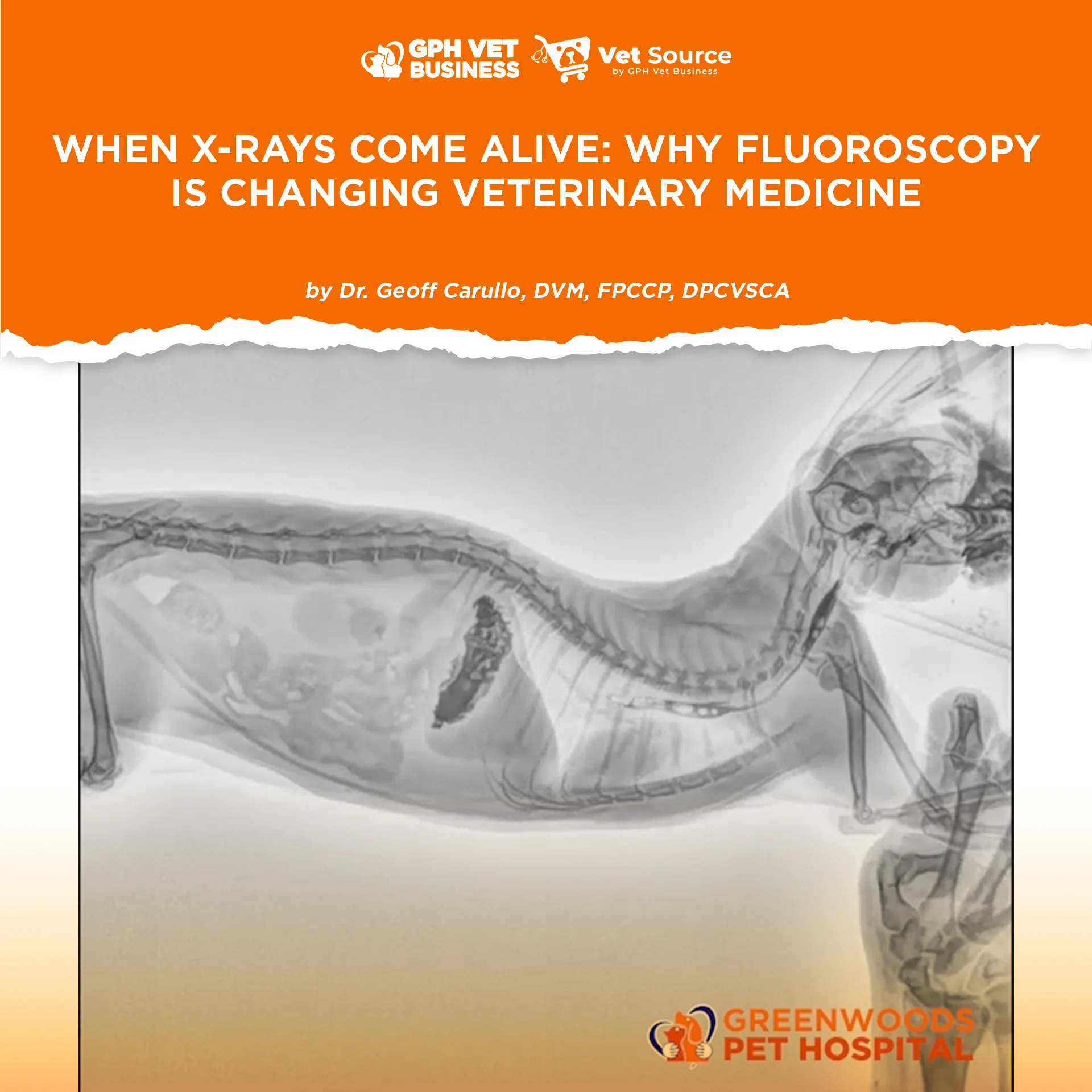
Figure 1. Fluoroscopic examination of a canine patient demonstrating the swallowing of Barium contrast in real time. This dynamic imaging technique allows veterinarians to observe the swallowing process and assess esophageal motility, helping identify potential swallowing disorders and functional abnormalities.
According to the University of California, Davis School of Veterinary Medicine, fluoroscopy is especially useful for diagnosing conditions involving movement and organ function.
WHY IS FLUOROSCOPY IMPORTANT IN SMALL ANIMAL PRACTICE?
Its clinical applications are extensive.
1. Detecting Dynamic Tracheal Collapse
Some dogs experience intermittent airway collapse that may not be fully appreciated on conventional radiographs.
Fluoroscopy allows veterinarians to evaluate changes in airway diameter during respiration.
This can be particularly valuable when assessing dogs with chronic coughing and suspected tracheal collapse.
2. Evaluating Swallowing Disorders
Figure 2. Real-time fluoroscopic examination demonstrating the movement of barium contrast within the stomach of a canine patient. This dynamic imaging technique allows veterinarians to evaluate gastric motility, observe contrast movement, and assess gastrointestinal function.
Patients suffering from regurgitation, dysphagia, or suspected esophageal dysfunction may benefit from fluoroscopic examination.
Using appropriate contrast studies, veterinarians can observe the movement of swallowed material through the esophagus and identify abnormalities in swallowing and transit.
3. Assessing Gastrointestinal Motility
Figure 3. Ventrodorsal fluoroscopic examination of a canine patient, demonstrating a prominent, oval radiopaque structure in the region of the urinary bladder. Real-time imaging allows veterinarians to assess its position and relationship to surrounding abdominal and pelvic structures, aiding further diagnostic evaluation.
Unlike conventional radiographs, fluoroscopy permits dynamic assessment of gastrointestinal movement.
It may help investigate selected swallowing and gastrointestinal motility disorders.
4. Guiding Minimally Invasive Procedures
Perhaps one of its most exciting applications is interventional radiology.
Veterinary specialists use fluoroscopic guidance during procedures involving:
- Tracheal and urethral stent placement
- Ureteral catheterization and stenting
- Balloon dilation of selected strictures
- Certain cardiovascular interventions
- Image-guided catheter and device placement
These techniques can sometimes reduce the need for more invasive surgical procedures.


DOES THIS MEAN CONVENTIONAL X-RAYS ARE BECOMING OBSOLETE?
Absolutely not.
Conventional digital radiography remains indispensable in veterinary practice.
Ultrasonography, computed tomography, and fluoroscopy each have their own advantages and limitations.
Fluoroscopy is particularly valuable when movement or real-time procedural guidance provides information that a static image cannot.
However, the technology requires significant investment, specialized training, appropriate facilities, and strict radiation safety protocols.
Because fluoroscopy uses ionizing radiation, veterinarians must follow the ALARA principle: As Low As Reasonably Achievable.
It is not simply a matter of buying an expensive machine.
Knowing when and how to use it is equally important.

THE FUTURE OF VETERINARY IMAGING IN THE PHILIPPINES
For many years, Philippine small animal practice relied heavily on conventional radiography and ultrasonography.
Today, veterinary medicine is gradually moving toward more sophisticated diagnostic and interventional technologies.
Fluoroscopy represents another opportunity to improve diagnostic capabilities, expand treatment options, and develop specialized veterinary services.
But advanced equipment should never be confused with advanced medicine.
A machine cannot replace clinical judgment, anatomical knowledge, experience, or proper case selection.
The real value of fluoroscopy is not that veterinarians can see more. It is that we can understand more about what is happening inside our patients.
And sometimes, that additional information can completely change a treatment plan.
Because in veterinary medicine, knowing what an organ looks like is important.
But knowing how it actually functions can be even more important.
SCIENTIFIC REFERENCES
UC Davis School of Veterinary Medicine. Fluoroscopy. Overview of diagnostic applications, including swallowing disorders, tracheal collapse, and gastrointestinal motility.
https://www.vetmed.ucdavis.edu/hospital/diagnostic-imaging/small-animal/fluoroscopy
UC Davis Veterinary Interventional Radiology. Overview of Veterinary Interventional Radiology. Applications of fluoroscopy in minimally invasive procedures.
https://ir.vetmed.ucdavis.edu/ir-applications/overview-veterinary-ir
University of Pennsylvania School of Veterinary Medicine. Interventional Radiology. Applications of image-guided veterinary procedures.
https://www.vet.upenn.edu/ryan-hospital/clinical-services/interventional-radiology/
American College of Veterinary Radiology. Radiation Safety Statement. Recommendations for minimizing occupational and patient radiation exposure.
https://acvr.org/how-we-do-it/types-of-imaging-therapy/radiology/acvrs-radiation-safety-statement/
These references support the diagnostic, interventional, and radiation-safety applications discussed above. (vetmed.ucdavis.edu)
Sharing this helps others understand what it really means to be a vet. Like and follow if you’re with us.
Dr. Geoff Carullo is a Fellow and the current President of the Philippine College of Canine Practitioners.