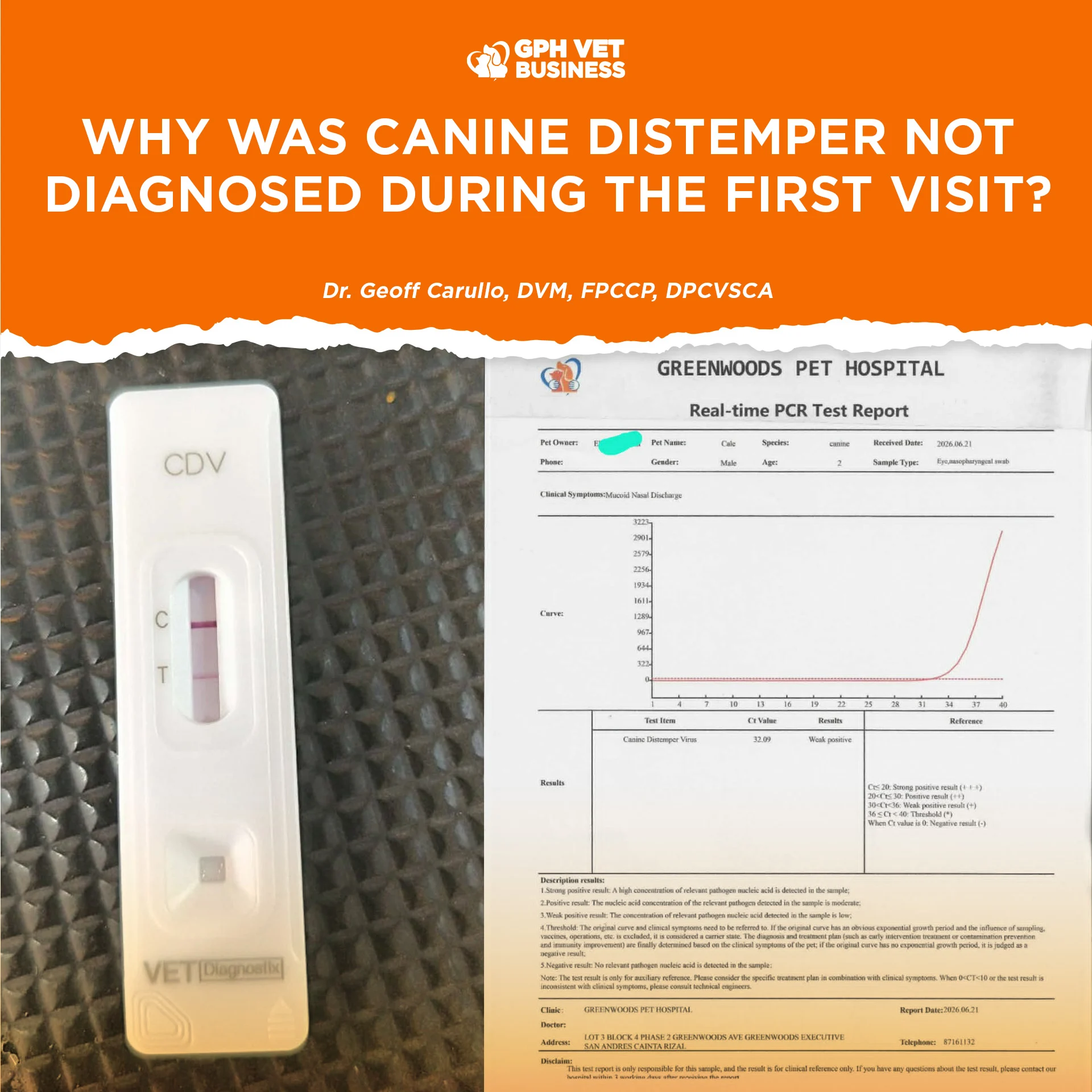
One of the most common misconceptions in veterinary medicine is the belief that every disease can be diagnosed during the first consultation.
Unfortunately, that is not how medicine works.
Recently, a patient was presented to Greenwoods Pet Hospital for chronic soft stool, poor appetite, and weight loss. During the initial consultation, the dog remained bright, alert, and responsive. There were no neurologic abnormalities. More importantly, there were no ocular discharges, no nasal discharges, and no respiratory signs that would immediately point toward canine distemper.
Based on the clinical findings available at that time, the patient was managed accordingly and scheduled for follow-up monitoring.
Several days later, the patient returned.
This time, the clinical picture had changed.
The dog had developed oculonasal discharge. Because of this new finding, canine distemper became a much stronger differential diagnosis. A canine distemper lateral flow test was performed and returned positive. PCR testing was subsequently conducted and also returned positive, confirming the diagnosis.
This naturally raises the question:
“Why wasn’t distemper diagnosed during the first visit?”
The answer is simple.
Because timing matters.
Canine distemper virus is a progressive disease. During the early stages of infection, many dogs exhibit vague and nonspecific signs such as fever, anorexia, lethargy, diarrhea, or weight loss. The more recognizable signs of distemper, including ocular discharge, nasal discharge, coughing, and neurological abnormalities, often appear later as the disease progresses.
In other words, the disease evolves.
Diagnostic tests are not magical tools that can detect every infection at every stage. They rely on the presence of detectable amounts of the pathogen in the sample collected.
For canine distemper, ocular and nasal secretions are among the most valuable samples for antigen detection and PCR testing. However, if a patient has not yet developed ocular or nasal discharge, the viral load within those sample sites may be low or even undetectable. Testing during this period may produce negative results despite the patient already being infected.
That is why veterinarians do not simply perform every possible test during the first consultation. Instead, diagnostic testing is guided by clinical findings, patient history, physical examination, and disease progression.


When the patient returned with oculonasal discharge, the situation changed dramatically.
The disease had progressed to a stage where viral detection became significantly more reliable. The clinical signs now matched the disease process, making both the lateral flow test and PCR testing highly valuable diagnostic tools.
Figure 1. Representative result of a canine distemper virus (CDV) lateral flow immunochromatographic assay. The presence of both the control (C) line and test (T) line indicates a positive result for canine distemper virus antigen. The visible control line confirms that the test functioned properly, while the test line demonstrates the detection of CDV antigen in the submitted sample. Positive results should always be interpreted in conjunction with the patient’s history, clinical signs, and other diagnostic findings such as hematology, PCR testing, and imaging studies.
The positive results obtained during the follow-up visit do not mean the disease suddenly appeared that day.
Figure 2. Real-time polymerase chain reaction (RT-PCR) amplification plot demonstrating the detection of canine distemper virus (CDV) nucleic acid from an eye and nasopharyngeal swab sample. The amplification curve exhibits a characteristic exponential rise crossing the detection threshold at a cycle threshold (Ct) value of 32.09, interpreted as a weak positive result, indicating the presence of a relatively low concentration of CDV genetic material in the specimen at the time of testing. This molecular finding confirms active viral infection and correlates with the patient’s clinical presentation of mucopurulent nasal discharge. PCR results should be interpreted alongside patient history, physical examination findings, and other diagnostic tests, including antigen-based lateral flow assays, to establish a comprehensive diagnosis.
Rather, it means the disease had progressed to the point where it could be detected more reliably.
This case highlights an important lesson for pet owners.
A Diagnosis Is Often a Journey, Not a Single Event
Veterinarians can only work with the information available at the time of examination. As new signs develop, diagnostic plans evolve. Follow-up consultations are not merely repeat visits. They are critical opportunities to reassess the patient, identify new findings, and arrive at a more accurate diagnosis.
Canine distemper is a disease that often reveals itself over time.
The absence of a diagnosis during the first visit does not necessarily mean the disease was overlooked.
Sometimes the disease simply has not shown its full story yet.
Veterinary medicine is not about guessing.
It is about performing the right test, on the right patient, at the right time.

References
- Cornell University College of Veterinary Medicine. Canine Distemper Virus.
- University of Georgia College of Veterinary Medicine. Ante-Mortem Diagnosis of Canine Distemper.
- Kansas State Veterinary Diagnostic Laboratory. Diagnostic Insights: Canine Distemper Virus Testing.
- Merck Veterinary Manual. Canine Distemper Virus Infection in Dogs.
- World Small Animal Veterinary Association (WSAVA) Vaccination Guidelines.
Sharing this helps others understand what it really means to be a vet. Like and follow if you’re with us.
Dr. Geoff Carullo is a Fellow and the current President of the Philippine College of Canine Practitioners.