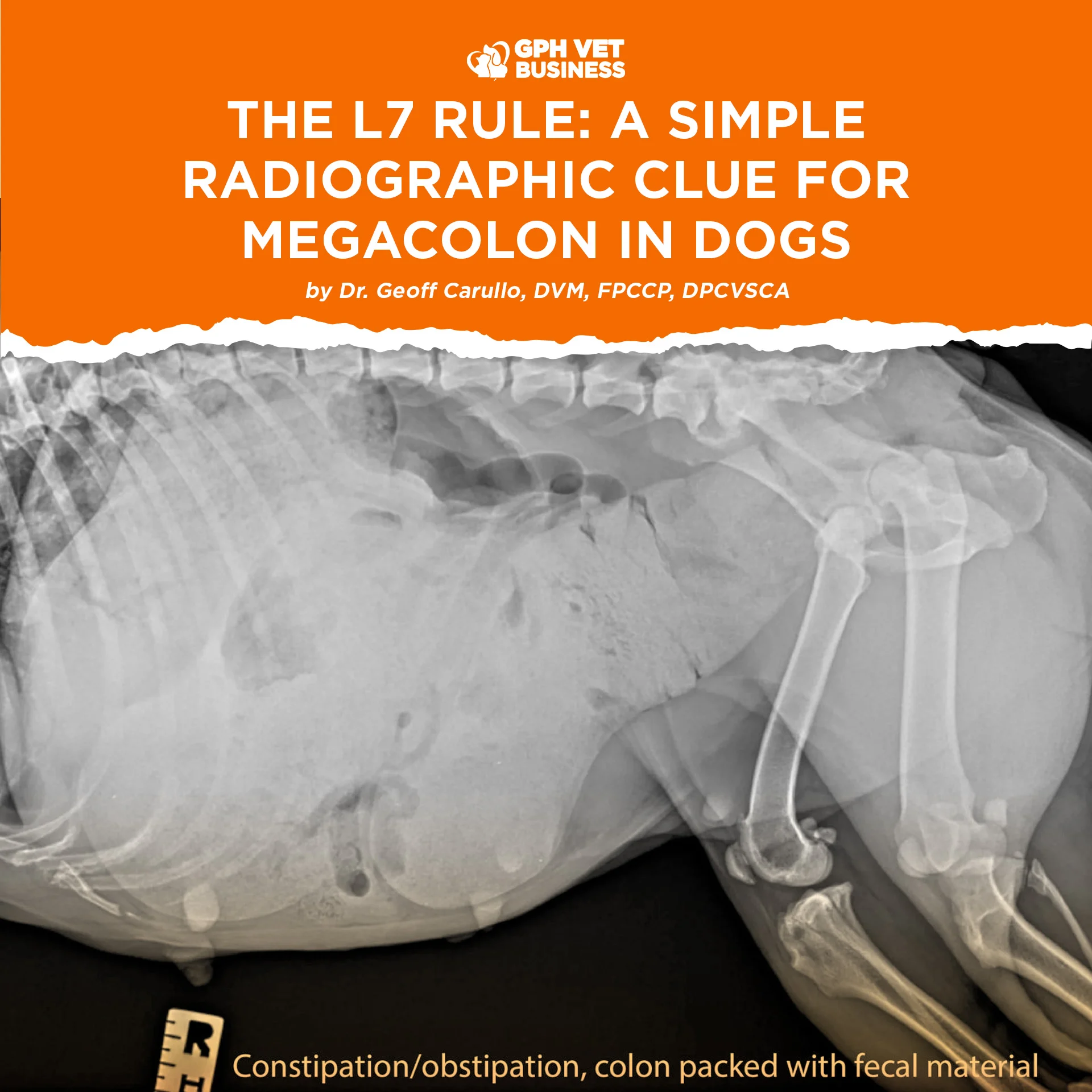
The L7 Rule: A Simple Radiographic Clue for Megacolon in Dogs
By Dr. Geoff Carullo, DVM, FPCCP, DPCVSCA
When a dog has been constipated for several days, seeing a colon packed with feces on an X-ray is easy. The more important question is:
Is this still constipation, or has the colon progressed to megacolon?
One useful radiographic clue is sitting right beside it: the seventh lumbar vertebra, or L7.


What Is Megacolon?
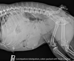
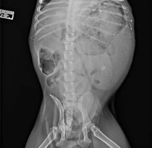
Megacolon refers to persistent, abnormal dilation of the colon associated with impaired colonic motility. Although it is much more commonly discussed in cats, dogs can develop megacolon as well.
In dogs, megacolon may develop secondary to chronic constipation or obstruction, pelvic canal narrowing, perineal hernia, colorectal disease, prostatic enlargement, neurologic dysfunction, or other conditions that interfere with normal defecation.
This is why simply seeing a large amount of feces on a radiograph is not enough.
Constipation describes difficulty passing feces. Megacolon implies significant dilation and dysfunction of the colon itself.
The L7 Rule
On a lateral abdominal radiograph, identify L7, the last lumbar vertebra immediately before the sacrum.
Measure two things:
- The length of the L7 vertebral body.
- The maximum diameter of the colon.
Then calculate:
MCD Ratio = Maximum Colonic Diameter ÷ L7 Vertebral Body Length
A commonly used radiographic guideline in dogs is:
MCD > 1.5 = strongly suggestive of megacolon
In practical terms, the maximal colonic diameter should generally not exceed approximately 1.5 times the length of the L7 vertebral body.
When the ratio exceeds this threshold, especially in a dog with chronic constipation or obstipation, the radiographic findings support the presence of megacolon.


A Simple Example
Suppose the L7 vertebral body measures 3.0 cm, while the maximum diameter of the colon measures 5.0 cm.
5.0 ÷ 3.0 = 1.67
Since 1.67 is greater than 1.5, the degree of colonic dilation would be strongly suggestive of megacolon when accompanied by compatible clinical findings.
Dogs and Cats Are Different
The vertebral reference used for assessing colonic dilation is not necessarily the same between dogs and cats.
For dogs, a practical radiographic guideline is:
Maximum colonic diameter ÷ L7 length > 1.5
For cats, a widely cited measurement uses L5, with a maximum colonic diameter-to-L5 ratio greater than approximately 1.48 supporting a diagnosis of megacolon.
This distinction is important. The feline L5 measurement should not simply be transferred to canine patients.
Do Not Diagnose Megacolon From the Ratio Alone
The L7 measurement is a radiographic guide, not a stand-alone diagnosis.
The measurement must always be interpreted together with the patient’s history, physical examination, clinical signs, and the remainder of the radiographic study.
Look for the complete clinical picture:
- Chronic or recurrent constipation or obstipation
- Repeated unsuccessful attempts to defecate
- Hard, dry feces
- Abdominal distension or discomfort
- A palpably enlarged, feces-filled colon
- Persistent marked colonic dilation on radiographs
- Poor response to conventional constipation management
Most importantly, look for the underlying cause.
Radiographs should also be evaluated for pelvic canal narrowing, previous pelvic fractures, masses, foreign material, perineal hernia, spinal abnormalities, prostatic enlargement, and other mechanical or neurologic causes of impaired defecation.
Depending on the patient, additional investigation may include bloodwork, rectal examination, ultrasonography, colonoscopy, CT, MRI, or neurologic evaluation.

The Take-Home Message
When evaluating a constipated dog, do not stop at:
“There is a lot of feces in the colon.”
Measure it.
The L7 vertebral body provides a convenient internal radiographic reference for assessing the severity of colonic dilation.
Maximum colonic diameter ÷ L7 length > 1.5? Think megacolon.
A large colon tells you that there is a problem. Comparing it with L7 helps objectively determine just how enlarged the colon has become.
And once megacolon is suspected, the next question should always be:
Why did the colon become this dilated in the first place?
Sources
Phansangiemjit A, et al. The Differences in Radiographic Vertebral Size in Dogs with Different Chest and Skull Types. Animals. 2024.
Use of Radiographic and Histologic Scores to Evaluate Cats With Idiopathic Megacolon Grouped Based on the Duration of Their Clinical Signs. Frontiers in Veterinary Science.
Türkiye Klinikleri. Megacolon and Its Surgical Treatment in Cats and Dogs.
Sharing this helps others understand what it really means to be a vet. Like and follow if you’re with us.
Dr. Geoff Carullo is a Fellow and the current President of the Philippine College of Canine Practitioners.