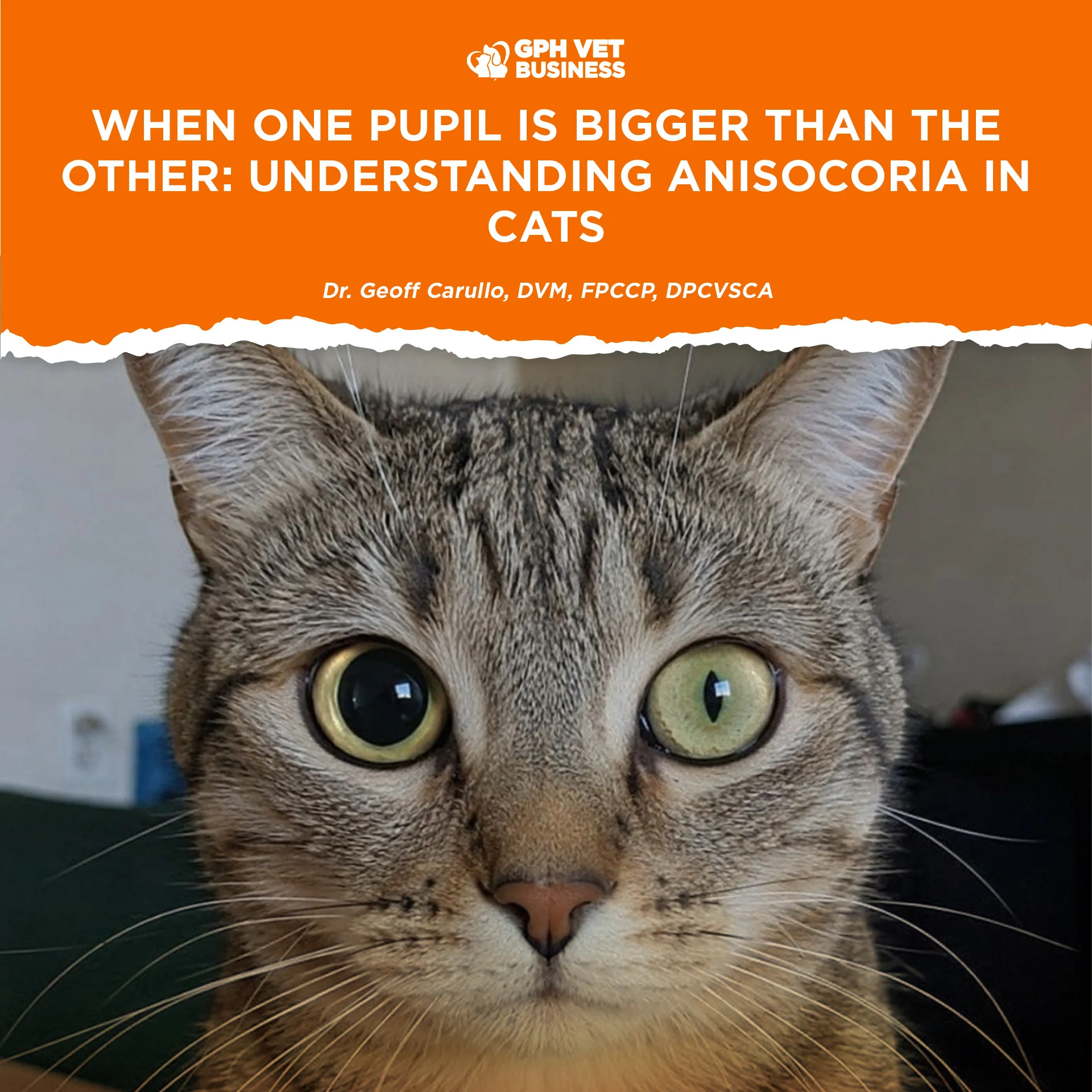
A cat with one pupil larger than the other may look fascinating, but from a veterinary standpoint, it should never be ignored.
This condition is called anisocoria, which simply means that the two pupils are unequal in size. It is not a disease by itself, but rather a clinical sign indicating that something is affecting the eye, the nerves controlling the eye, or even the brain.
Some causes are relatively minor.
Others can threaten vision, or even the life of the patient.
That is why anisocoria should always be approached as a diagnostic challenge rather than dismissed as “just different pupils.”
What Controls Pupil Size?
The pupil constantly changes size to regulate how much light enters the eye.
This process is controlled by two opposing parts of the autonomic nervous system:
- Parasympathetic system – constricts the pupil (miosis)
- Sympathetic system – dilates the pupil (mydriasis)
Damage to either pathway can result in one pupil becoming larger or smaller than the other.


Common Causes of Anisocoria in Cats
1. Horner’s Syndrome
One of the most recognizable neurological causes.
Clinical signs include:
- Small pupil (miosis)
- Elevated third eyelid
- Slight drooping of the upper eyelid (ptosis)
- Sunken appearance of the eye (enophthalmos)
Horner’s syndrome develops when the sympathetic nerve supply to the eye is interrupted.
Possible causes include:
- Ear disease
- Neck trauma
- Bite wounds
- Chest lesions
- Brain disease
- Idiopathic cases
2. Anterior Uveitis
Inflammation inside the eye commonly causes:
- Pain
- Red eye
- Excessive tearing
- Cloudiness
- Constricted pupil
Cats with uveitis require prompt investigation because underlying causes may include:
- Feline infectious diseases
- Trauma
- Immune-mediated disease
- Neoplasia
Failure to treat may result in glaucoma or permanent blindness.
3. Glaucoma
Increased intraocular pressure damages the optic nerve.
Typical findings include:
- Enlarged pupil
- Painful eye
- Corneal edema
- Vision loss
Glaucoma is considered an ophthalmic emergency because irreversible retinal damage can occur within a short period if pressure remains elevated.
4. Corneal Ulcers
Pain from a corneal ulcer can produce reflex miosis.
These cats often present with:
- Squinting
- Excessive tearing
- Rubbing the eye
- Photophobia
Fluorescein staining is essential before prescribing topical corticosteroids.


5. Trauma
Head injuries can damage:
- Iris muscles
- Cranial nerves
- Sympathetic nerves
- Globe structures
Traumatic anisocoria should never be underestimated, especially following vehicular accidents or falls.
6. Retinal Disease or Blindness
If one eye loses vision because of retinal disease or optic nerve damage, the affected pupil may fail to constrict normally when exposed to light.
This is why assessing vision and pupillary light reflexes is critical during examination.
7. Neurologic Disease
Brain lesions affecting cranial nerves III or sympathetic pathways may produce anisocoria.
Possible causes include:
- Brain tumors
- Encephalitis
- Stroke
- Intracranial trauma
A complete neurological examination becomes essential whenever ocular findings are accompanied by changes in mentation, gait abnormalities, seizures, or cranial nerve deficits.
Diagnosing Anisocoria
The pupils alone never provide the diagnosis.
Instead, they provide an important clue.
A complete work-up may include:
- Full ophthalmic examination
- Menace response
- Dazzle reflex
- Pupillary light reflex testing
- Schirmer tear test
- Fluorescein staining
- Tonometry (measurement of intraocular pressure)
- Slit-lamp biomicroscopy
- Fundic examination
- Neurologic examination
- Blood work
- Blood pressure measurement
- Imaging such as radiographs, CT, or MRI when indicated
Veterinarians must determine which pupil is abnormal—the one that is too small or the one that is too large. That distinction significantly narrows the list of differential diagnoses.

Is It an Emergency?
Yes.
Sudden-onset anisocoria should always be considered urgent until proven otherwise.
Delaying evaluation may result in:
- Permanent blindness
- Glaucoma progression
- Untreated neurological disease
- Missed ocular emergencies
The earlier the diagnosis, the greater the chance of preserving vision.
Final Thoughts
Anisocoria is one of those findings that reminds us why a careful physical examination still matters.
Two pupils that are different sizes may be the first visible sign of ocular inflammation, glaucoma, Horner’s syndrome, retinal disease, trauma, or serious neurological disorders.
The unequal pupils themselves are not the disease.
They are the body’s way of telling us that something deeper deserves our attention.
For veterinarians, anisocoria should never be ignored.
For pet owners, it should never be treated as “normal.”
Prompt examination can make the difference between preserving vision and losing it forever.
References
- Zwueste DM, Giuliano EA. A Review of Horner’s Syndrome in Small Animals. Canadian Veterinary Journal. 2019.
- VCA Animal Hospitals. Anisocoria in Cats.
- Today’s Veterinary Practice. The Practitioner’s Guide to Neurologic Causes of Anisocoria.
- Clinician’s Brief. Elevation of the Third Eyelid and Miosis in a Cat: Horner’s Syndrome.
- PetMD. Anisocoria in Cats.
- PetMD. Horner’s Syndrome in Cats.
Dr. Geoff Carullo is a Fellow and the current President of the Philippine College of Canine Practitioners.
Sharing this helps others understand what it really means to be a vet. Like and follow if you’re with us.