A dog with urinary stones can present with anything from mild hematuria to life-threatening acute kidney injury. The challenge for veterinarians is not simply identifying the presence of stones, but determining their exact location, size, number, and whether they are causing obstruction.
The combination of radiography and ultrasonography remains one of the most powerful diagnostic approaches for canine urolithiasis.
The images shown in this case demonstrate why both modalities are often necessary.
What Radiography Can Tell Us
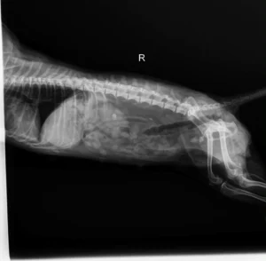
Figure 1. Right lateral abdominal radiograph of a dog demonstrating multiple radiopaque urinary calculi. Mineral-dense opacities are visible in the regions of both kidneys, with additional calculi noted along the urinary tract, consistent with multifocal urolithiasis involving the kidneys, ureters, and urinary bladder. Radiography provides an excellent initial assessment of stone number, size, and distribution within the urinary system.
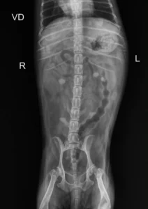
Figure 2. Ventrodorsal abdominal radiograph demonstrating bilateral nephrolithiasis. Multiple well-defined radiopaque calculi are visible within the anatomical regions of both kidneys. Additional mineral opacities are present along the urinary tract, supporting the diagnosis of multifocal urolithiasis. The ventrodorsal projection provides excellent visualization of stone distribution and aids in assessing the number, size, and laterality of urinary calculi.
Survey radiographs remain the first-line imaging tool for many urinary stone cases.
In this patient, the abdominal radiographs reveal multiple mineral-dense structures within the urinary tract. Radiopaque calculi are visible in the region of both kidneys and along the urinary tract.
Radiography is particularly useful for:
- Detecting radiopaque stones such as struvite, calcium oxalate, calcium phosphate, and silica uroliths
- Determining the number and approximate size of stones
- Monitoring changes over time
- Assessing treatment response
- Screening for recurrent stone formation
Radiographs are highly sensitive for mineralized calculi but may miss radiolucent stones such as urate and cystine uroliths.


What Ultrasonography Adds
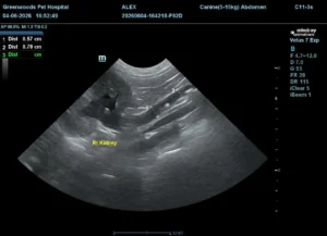
Figure 3. Ultrasonographic image of the right kidney demonstrating a dilated ureter containing a hyperechoic ureterolith measuring approximately 0.57 to 0.70 cm. The mineralized structure produces distal acoustic shadowing and is associated with hydroureter, consistent with obstructive ureterolithiasis. Ultrasonography is particularly valuable for identifying ureteral calculi and evaluating the degree of urinary tract obstruction.
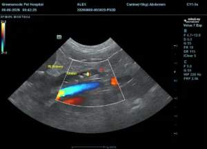
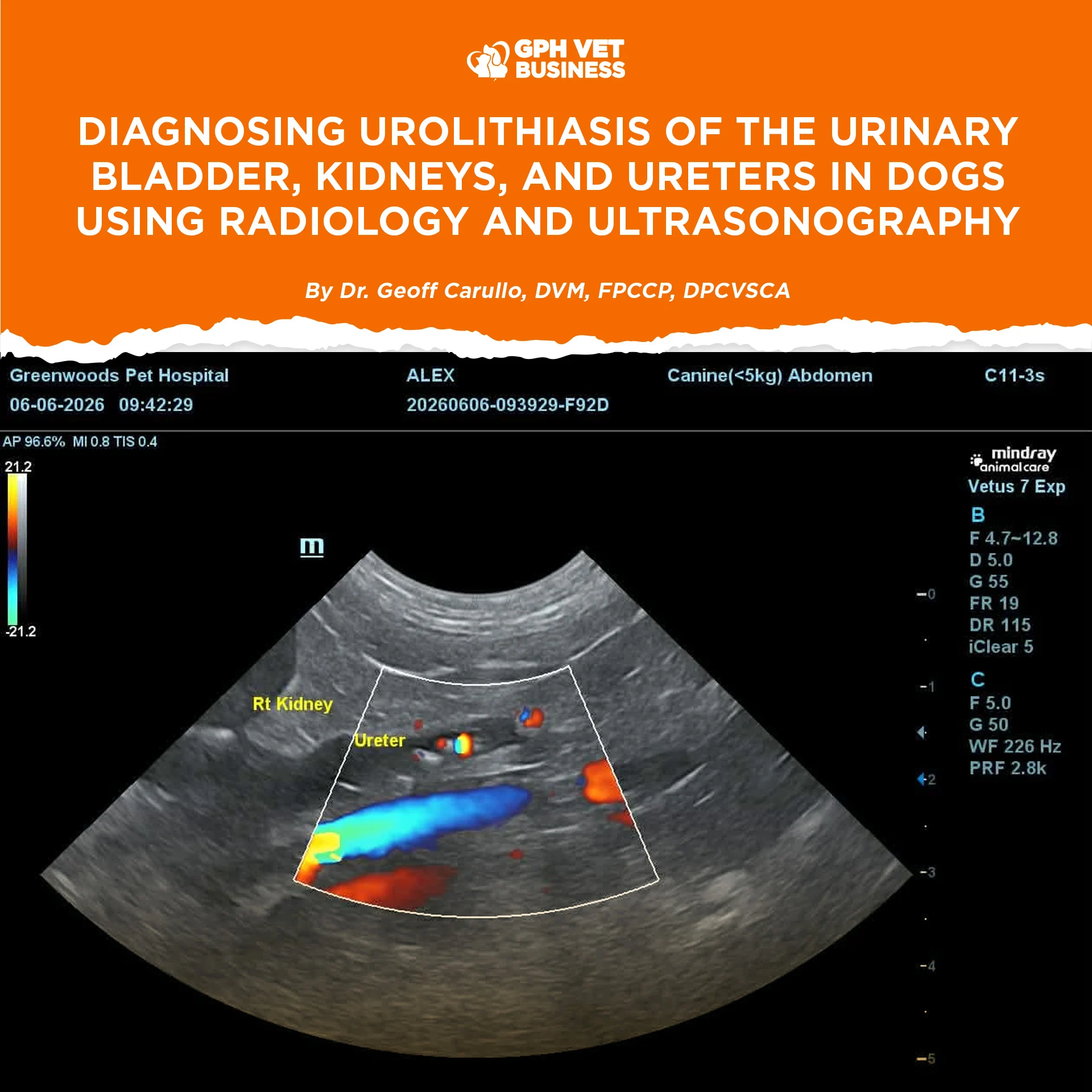
Figure 4. Ultrasonographic image of the right kidney and proximal ureter in a dog. The ureter is visualized as a tubular anechoic structure adjacent to the right kidney. Color Doppler interrogation confirms the presence of blood flow in adjacent vessels and assists in differentiating the ureter from surrounding vascular structures. Ureteral dilation (hydroureter) may be observed in cases of ureterolithiasis causing partial or complete urinary obstruction. The absence of Doppler flow within the ureter helps distinguish it from nearby blood vessels.
While radiographs identify mineral opacities, ultrasonography provides information about soft tissues and urinary tract function.
In this patient, ultrasound images demonstrate:
- Renal calculi within the kidney
- Ureteral dilation
- Mineralized structures within the ureter
- Evidence suggestive of ureterolithiasis
The ultrasonographic appearance of ureteral obstruction typically includes:
- Hyperechoic mineralized focus
- Distal acoustic shadowing
- Proximal ureteral dilation
- Renal pelvic dilation (pyelectasia)
- Hydronephrosis in severe cases
Color Doppler can further assist in identifying vascular structures and differentiating vessels from dilated ureters.
Why Ureteral Stones Matter
Many bladder stones cause discomfort.
Ureteral stones can destroy kidneys.
A ureterolith may obstruct urine flow from the kidney to the bladder. Even partial obstruction can lead to progressive renal damage.
Common findings include:
- Hydroureter
- Hydronephrosis
- Renal pelvic dilation
- Azotemia
- Acute kidney injury
Dogs with bilateral ureteral obstruction may rapidly become critical patients.

The Power of Combining Both Modalities
Neither radiography nor ultrasonography should be considered sufficient in every case.
Radiography answers:
“Where are the stones?”
Ultrasound answers:
“What are those stones doing to the urinary tract?”
Together they allow clinicians to:
- Confirm nephroliths
- Confirm ureteroliths
- Identify cystoliths
- Assess obstruction severity
- Evaluate renal damage
- Guide treatment planning
This multimodal approach often determines whether a patient requires medical management, cystotomy, ureterotomy, ureteral stenting, SUB placement, or referral for advanced intervention.
Clinical Take-Home Message
When evaluating dogs with hematuria, dysuria, pollakiuria, recurrent urinary tract infections, or unexplained azotemia, always remember that stones may exist in multiple locations simultaneously.
A bladder stone may be the most obvious finding.
The kidney stone may be the most important finding.
But the ureteral stone may be the one that ultimately threatens the patient’s life.
Radiographs help us see the stones.
Ultrasound helps us understand their consequences.
Using both imaging modalities together gives veterinarians the best opportunity to accurately diagnose and successfully manage canine urolithiasis.
References
BSAVA Manual of Canine and Feline Nephrology and Urology
American College of Veterinary Internal Medicine Consensus Recommendations for Management of Urolithiasis in Dogs and Cats.
International Renal Interest Society Guidelines on Obstructive Uropathy and Acute Kidney Injury.
Berent AC. Ureteral Obstruction in Dogs and Cats: Diagnosis and Interventional Management. Veterinary Clinics of North America: Small Animal Practice.
Nyland TG, Mattoon JS. Small Animal Diagnostic Ultrasound.
Thrall DE. Textbook of Veterinary Diagnostic Radiology.
Dr. Geoff Carullo is a Fellow and the current President of the Philippine College of Canine Practitioners.
Sharing this helps others understand what it really means to be a vet. Like and follow if you’re with us.