There’s a moment every veterinarian recognizes.
You’re reviewing a late-term pregnancy X-ray.
Most fetuses look normal.
Then one image stops you.
Something looks… wrong.
Not distorted.
Not gassy.
Just collapsed.
That’s when mummified fetus should immediately enter your differential.
What Is a Mummified Fetus (Radiographically Speaking)?
A mummified fetus is a fetus that died in utero, remained sterile, and slowly dehydrated instead of decomposing.
No bacteria.
No gas.
No maceration.
Just a dry, shrunken fetus trapped inside the uterus.
Radiographs are often the first clue.
Key X-ray Signs of a Mummified Fetus
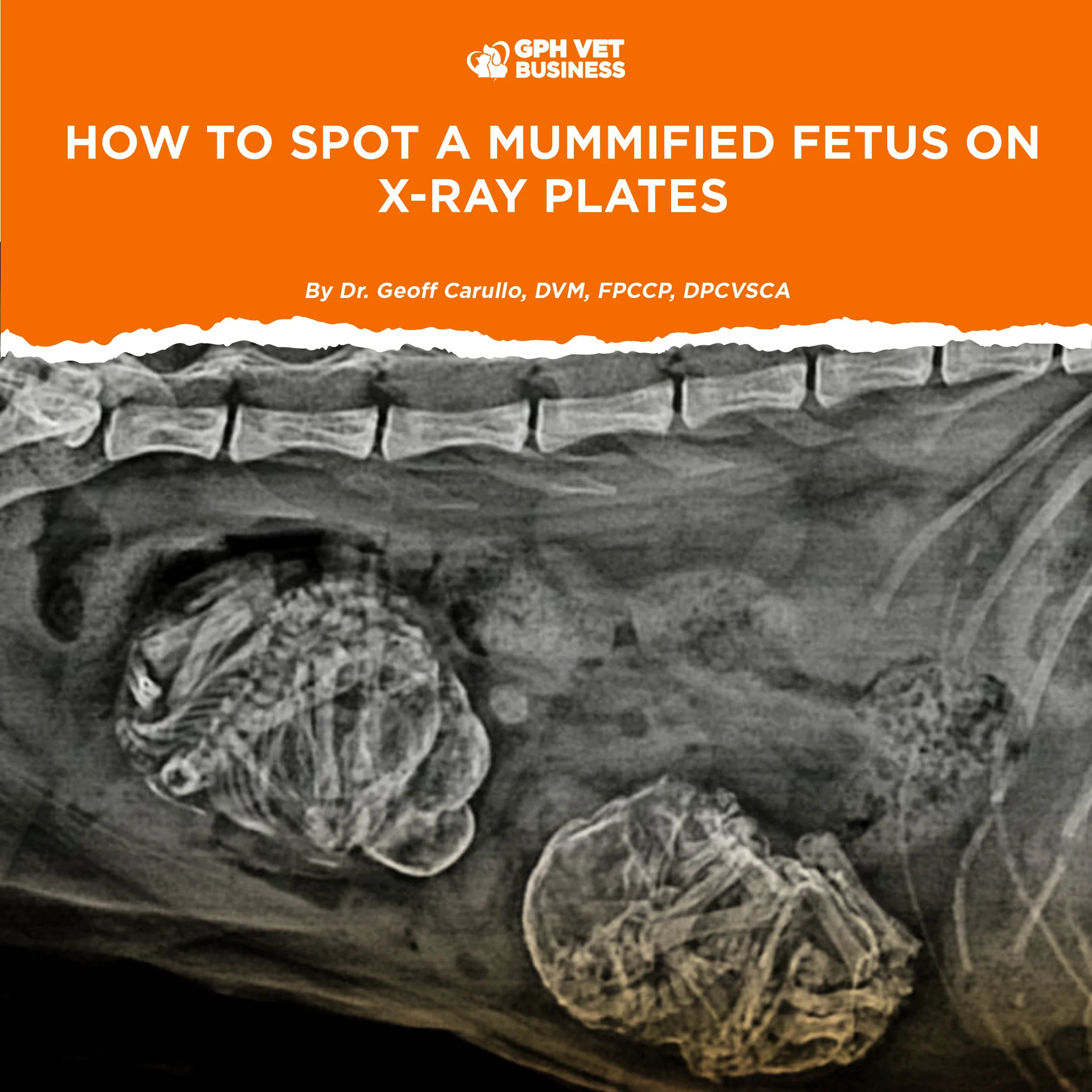
1. Collapsed and Overlapping Skeleton
This is the most reliable sign.
On X-ray, you’ll see:
- Bones folded onto each other
- Loss of normal fetal posture
- Skull, spine, ribs overlapping unnaturally
A healthy fetus looks organized.
A mummified fetus looks crumpled.
If it resembles a “pile of bones” instead of a fetus — pay attention.
2. Smaller Than Expected for Gestational Age
In late pregnancy, all fetuses should be roughly similar in size.
A red flag appears when:
- One fetus is significantly smaller
- Others are full-term size
A fetus that dies stops growing immediately.
The uterus keeps going — the fetus doesn’t.
3. Sharp, Dry Bone Margins
Mummification removes fluid and soft tissue.
Radiographically this appears as:
- Very clear and sharp bone outlines
- Minimal to absent soft tissue shadow
The fetus looks “too skeletal” for its age.
4. Absence of Gas (Very Important)
This helps you differentiate conditions.
Mummified fetus:
No gas. No bloating.
Macerated fetus:
Gas present. Bones fragmented.
No gas means:
- Sterile uterine environment
- Closed cervix
- Classic mummification
5. Isolated Among Normal Fetuses
Most cases are partial litter loss.
Common scenario:
- One abnormal fetus
- Remaining fetuses appear normal
This often delays diagnosis because labor may still start — but not progress.
6. No Change on Repeat X-rays
If serial radiographs are taken:
- Viable fetuses may shift position
- A mummified fetus stays completely stationary
Dead, dehydrated tissue does not move.
Quick Radiographic Checklist
If you see three or more of the following, strongly suspect mummification:
- ✔ Collapsed fetal skeleton
- ✔ Overlapping bones
- ✔ Smaller than littermates
- ✔ Sharp bone margins
- ✔ Minimal soft tissue
- ✔ No gas
- ✔ No positional change over time
Why This Matters Clinically
A mummified fetus can:
- Block the birth canal
- Prevent proper uterine contractions
- Cause prolonged gestation
- Lead to dystocia despite normal fetuses
This is why many of these cases end in cesarean section, even when other pups are viable.
Waiting too long often risks:
- Fetal loss
- Uterine fatigue
- Maternal complications
Final Clinical Pearl
Radiographs don’t just confirm pregnancy.
They tell a story.
When one fetus looks dry, collapsed, and frozen in time —
that story is mummification.
Spot it early.
Plan decisively.
Save the bitch — and the remaining litter.
Sharing this helps others understand what it really means to be a vet. Like and follow if you’re with us.