Ultrasound assessment of the kidneys is one of the most common—and most powerful—applications of caliper measurement in small animal practice. Beyond a subjective “normal vs abnormal” impression, objective renal measurements allow veterinarians to distinguish acute from chronic disease, identify obstruction early, and monitor progression or response to therapy with confidence.
This article outlines the essential kidney caliper measurements every clinician should routinely document during abdominal ultrasound.
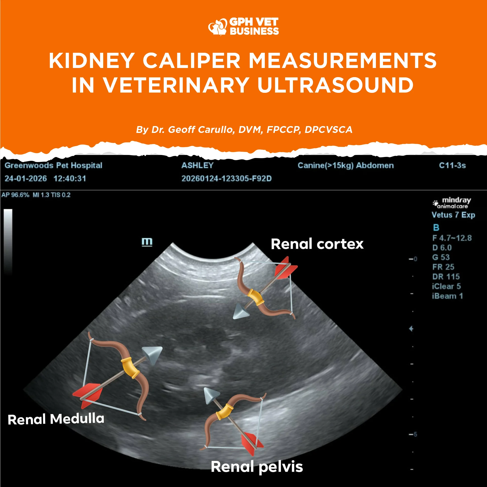
Renal Anatomy: What You Must Identify First
Accurate measurements start with correct anatomical orientation on ultrasound:
- Renal capsule – thin, hyperechoic outer line
- Renal cortex – moderately hypoechoic outer parenchyma
- Renal medulla – hypoechoic to anechoic pyramids
- Renal pelvis – central echogenic region, sometimes with anechoic fluid
Misidentifying these structures leads to incorrect caliper placement and misleading conclusions.
1. Kidney Length (Primary and Most Important Measurement)
How to measure
- Use a longitudinal view
- Measure from outer cranial pole to outer caudal pole
- Place calipers along the true long axis (not curved)
Normal reference
- Dogs: ~2.5–3.5 × the length of the L2 vertebra
- Cats: approximately 3.0–4.3 cm in most adult cats
Clinical significance
- Enlarged kidneys → acute kidney injury, pyelonephritis, lymphoma
- Small kidneys → chronic kidney disease (irreversible change)
Kidney length is the single most reliable objective indicator of renal size and chronicity.
2. Cortical Thickness
How to measure
- Measure from the renal capsule to the corticomedullary junction
- Best assessed at the mid-kidney region
Typical values
- Dogs: ~5–8 mm (size dependent)
- Cats: ~4–6 mm
Interpretation
- Thickened cortex suggests acute inflammation or edema
- Thinned cortex is strongly associated with chronic renal disease
3. Medullary Thickness
How to measure
- From the corticomedullary junction to the renal pelvis
Clinical notes
- Medulla should be similar to or slightly thicker than the cortex
- Loss of normal proportions or corticomedullary distinction indicates advanced renal pathology
4. Renal Pelvis Diameter
How to measure
- Measure the widest anechoic portion of the renal pelvis on longitudinal view
Normal
- ≤2–3 mm in dogs and cats
Abnormal
- 4 mm suggests pyelectasia
- Marked dilation indicates obstruction or hydronephrosis
- Mild pelvic dilation can be physiologic after IV fluids—always interpret with clinical context.
5. Renal Width and Height (Adjunct Measurements)
Measured in transverse view, these are helpful for:
- Comparing symmetry between kidneys
- Monitoring renal masses or cysts
- Serial follow-up examinations
They are supportive but never replace kidney length.
6. Renal Masses and Cysts
When present:
- Measure in at least two planes
- Record length × width (± height)
- Document location (cortical, medullary, pelvic), margins, and echogenicity
Consistent measurements are essential for monitoring growth or treatment response.
Sample Ultrasound Report Entry
Right kidney measures 6.8 cm in length with cortical thickness of 6 mm and renal pelvis diameter of 1.5 mm.
Left kidney measures 6.5 cm with cortical thickness of 5.5 mm and pelvis diameter of 1.3 mm.
Corticomedullary distinction is preserved. No evidence of pyelectasia.
Key Takeaways for Clinical Practice
- Kidney length is the cornerstone of renal ultrasound evaluation
- Cortical and pelvic measurements provide insight into disease stage and chronicity
- Always compare left vs right kidneys
- Correlate ultrasound findings with creatinine, SDMA, and urinalysis
- Objective caliper measurements transform renal ultrasound from a descriptive tool into a decisive diagnostic instrument.
Sharing this helps others understand what it really means to be a vet. Like and follow if you’re with us.